Atherectomy Explained: What It Is, Why It’s Done, and What to Expect?
- Dhanvin Raj Puppala
- Mar 26
- 5 min read


What it is?
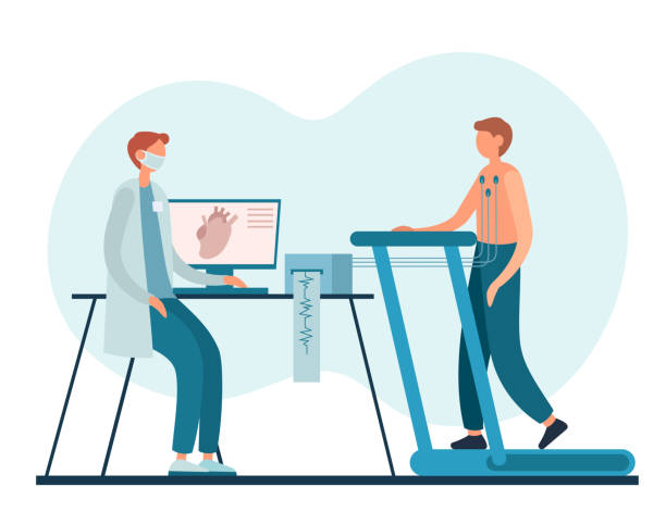
Atherectomy is a catheter based procedure used to remove or modify plaque inside an artery. Plaque is a buildup of cholesterol, calcium, and scar like material that can narrow blood vessels and reduce blood flow. Atherectomy is performed through a small tube placed into an artery, often in the groin or wrist, and is guided by X ray imaging.¹,²
Atherectomy is one tool in a larger set of treatments for blocked arteries. It is commonly used as “vessel preparation,” meaning it helps open a tight or heavily calcified blockage so that other treatments, like balloon angioplasty, drug coated balloons, or stents, can work better.²
Where it is used
Atherectomy is used in two main settings:
Leg arteries (peripheral artery disease, PAD)PAD can cause leg pain with walking (claudication) or, in severe cases, chronic limb threatening ischemia, where poor blood flow contributes to nonhealing wounds, tissue loss, or rest pain. Guideline based PAD care emphasizes risk factor treatment (for example, smoking cessation, cholesterol lowering, blood pressure control, diabetes management), exercise therapy, and selected cases of revascularization when symptoms are lifestyle limiting or limb threatening.¹
Heart arteries (coronary artery disease)In the heart, calcium can make it hard to expand balloons and fully open a stent. Expert consensus documents describe atherectomy and other calcium modification methods as options when imaging or angiography shows severe calcification that would likely prevent proper stent expansion.²
Types of atherectomy in simple terms
Different devices remove plaque in different ways. Your clinician chooses based on artery size, plaque type, calcium amount, and location.²,⁷
Directional atherectomy: shaves plaque from one side of the vessel and collects it in a nosecone on the device.³,⁴
Rotational or orbital atherectomy: sands or ablates calcified plaque, mainly used for calcium modification.²
Laser atherectomy: uses laser energy to break down plaque, sometimes used in complex leg artery disease.⁶
Potential benefits
Atherectomy may help in specific situations, especially when plaque is bulky or calcified.
It can improve the immediate opening of the artery and reduce the need for emergency stenting in some scenarios by making the vessel more responsive to balloon treatment.⁴
In a large prospective study of directional atherectomy in leg arteries, outcomes at 12 months suggested it can be effective in appropriately selected patients, with a relatively low rate of bailout stenting reported in that study.³
In a randomized pilot study comparing directional atherectomy plus a drug coated balloon versus drug coated balloon alone in femoropopliteal disease, technical success was higher and flow limiting dissections were lower in the combined group, while one year clinical outcomes were similar in this underpowered trial.⁴
Important limitations and ongoing debate
A key point for the public to understand is that “more technology” does not automatically mean “better outcomes.” Evidence for atherectomy includes randomized trials, registries, and observational studies, but comparative data are not always definitive across all patient groups and lesion types.³,⁴,⁷
Real world practice variation has also raised concerns about overuse in some settings. A Medicare claims analysis found wide differences among physicians in how often atherectomy was used for femoropopliteal interventions and highlighted the need for clear appropriate use standards.⁵
A broad systematic review and meta analysis reported generally favorable average outcomes across many atherectomy studies, but also emphasized high heterogeneity and the need for stronger comparative research.⁷
Risks and side effects
All catheter based artery procedures carry risks. With atherectomy, common concerns include:
Distal embolization: small debris can travel downstream and block smaller vessels, sometimes requiring additional treatment.³,⁷
Artery injury: including dissection (a tear in the artery lining) or perforation.³
Bleeding or bruising at the access site, infection, allergic reaction to contrast dye, and kidney stress from contrast in susceptible patients.¹,³
Need for repeat procedures if the artery narrows again over time (restenosis).⁴,⁶
Your personal risk depends on overall health, kidney function, diabetes control, the length and location of the blockage, and whether the disease is limb threatening.¹,⁶
Questions to ask your clinician
If atherectomy is being recommended, consider asking:
What is the goal in my case: symptom relief, wound healing, or limb salvage?¹
What are the alternatives: supervised exercise therapy, medication optimization, balloon angioplasty, drug coated balloon, stent, bypass surgery, or no procedure?¹
What specific device type will be used and why?²
Will you use imaging inside the artery to assess calcium and result quality?²
What are the risks of embolization, vessel injury, and need for additional stents in my anatomy?³,⁴
What follow up is planned and what medications will I need afterward?¹
Conclussion
Atherectomy is a specialized technique to remove or modify plaque, most often used to prepare difficult blockages, especially those with heavy calcium, for other treatments.² It can be helpful for selected patients, but it is not automatically the best choice for everyone, and the strongest evidence supports careful patient selection and attention to overall guideline based PAD and cardiovascular care.¹,⁵,⁷
Citation
Gornik HL, Aronow HD, Goodney PP, Arya S, Brewster LP, Byrd L, et al. 2024 ACC/AHA multisociety guideline for the management of lower extremity peripheral artery disease. Circulation. 2024;149(24):e1313–e1410.
Riley RF, Patel MP, Abbott JD, Bangalore S, Brilakis ES, Croce KJ, et al. SCAI expert consensus statement on the management of calcified coronary lesions. J Soc Cardiovasc Angiogr Interv. 2024;3(2):101259.
McKinsey JF, Zeller T, Rocha-Singh KJ, Jaff MR, Garcia LA; DEFINITIVE LE Investigators. Lower extremity revascularization using directional atherectomy: 12-month results of the DEFINITIVE LE study. JACC Cardiovasc Interv. 2014;7(8):923-933.
Zeller T, Langhoff R, Rocha-Singh KJ, Jaff MR, Blessing E, Amann-Vesti B, et al. Directional atherectomy followed by a paclitaxel-coated balloon to inhibit restenosis and maintain vessel patency: 12-month results of the DEFINITIVE AR study. Circ Cardiovasc Interv. 2017;10(9):e004848.
Hicks CW, Holscher CM, Wang P, Dun C, Abularrage CJ, Black JH III, et al. Use of atherectomy during index peripheral vascular interventions. JACC Cardiovasc Interv. 2021;14(6):678-688.
Stoner MC, deFreitas DJ, Phade SV, Parker FM, Bogey WM, Powell S. Mid-term results with laser atherectomy in the treatment of infrainguinal occlusive disease. J Vasc Surg. 2007;46(2):289-295.
Chambers JW, Diage T, McGuffin M, Patel M, McEntegart M. Outcomes of rotational atherectomy in heavily calcified coronary lesions. Catheter Cardiovasc Interv. 2014;83(3):E141-E148.
Carr JG, Langhoff R, DeRubertis BG, Hood KL, Krishnan P, Puttaswamy V, et al. Published evidence on peripheral atherectomy: a systematic review and meta-analysis. J Soc Cardiovasc Angiogr Interv. 2025;4(11):104009.
Image:Willis-Knighton Health. (n.d.). Atherectomy (Rotational) [Illustration]. Retrieved January 21, 2026, from the Willis-Knighton Health website.
Assessed and Endorsed by the MedReport Medical Review Board